By Connie Chen, Microbiology, ‘16
Author’s Note:
“Many areas of employment, especially within health care, require employees to take a test to see if they have been exposed to tuberculosis (TB). Today, it is believed that one third of the world’s population is infected with some form of TB. However, not many people truly understand what tuberculosis is or what it does. I hope that after reading this, you will have a better understanding of TB.”
Pathogenesis:
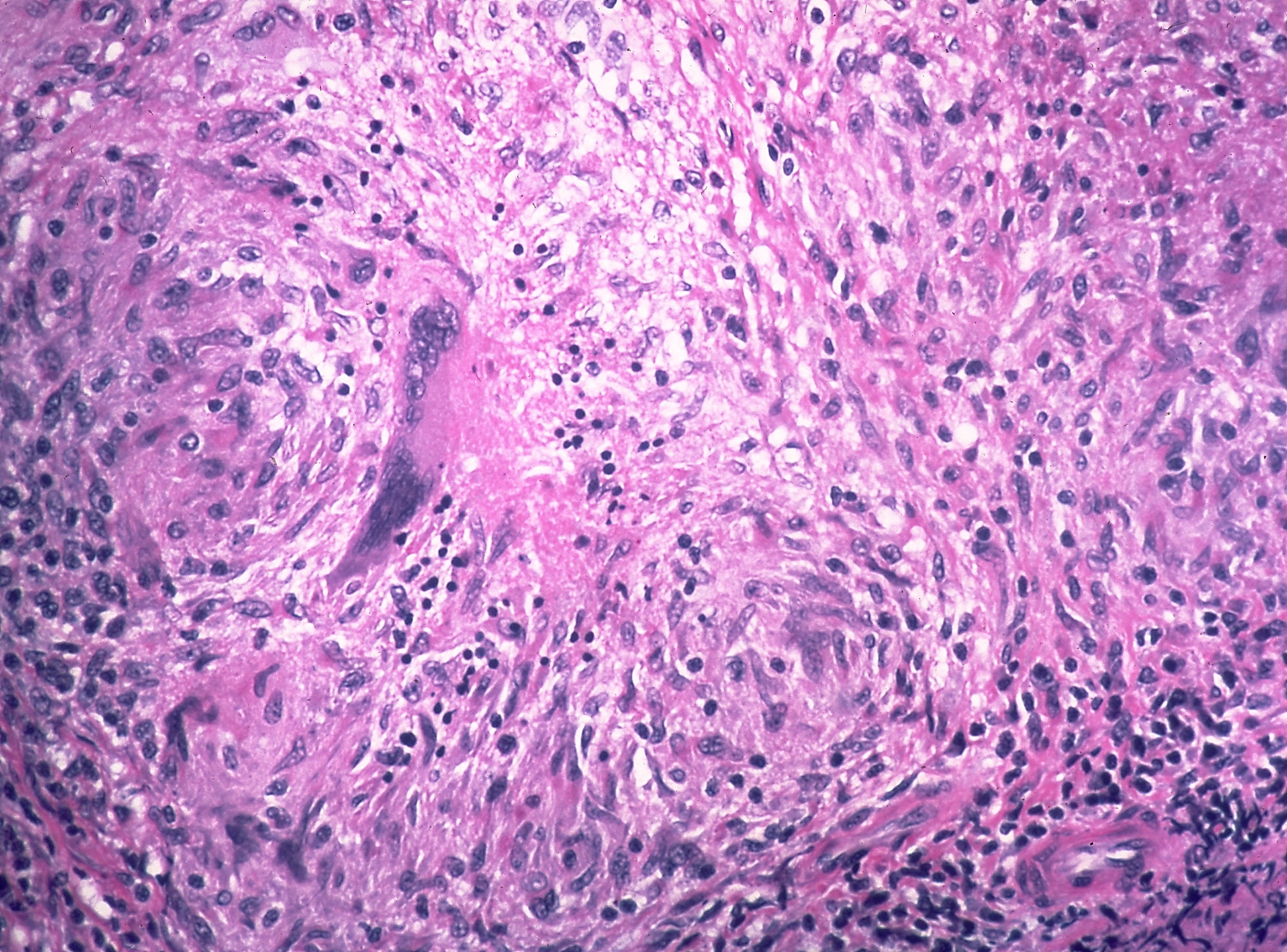
When TB was an epidemic in the United States during the early 1900’s, many people nicknamed the disease “consumption” because of how it “eats you away.” For those with active TB, some symptoms include fever, chills, night sweats and weight loss. Most cases of tuberculosis are caused by the bacterium, Mycobacterium tuberculosis. Although there are other Mycobacterium species that cause TB, M. tuberculosis is the main causative agent. M. tuberculosis has many unique characteristics that make it stand out. A notable feature is the bacterium’s thick, waxy, mycolic acid coating, which plays a vital role in its survival within the human body. Although most bacteria can survive for a few days on dry surfaces such as a tabletop, the waxy coat allows M. tuberculosis to survive for weeks in a dry state. This also helps increase resistance to dehydration and chemical damage, which can prevent antibiotics from killing the organism (Lampart, 2002).
- M. tuberculosis prefers to colonize in the lungs because it is highly aerobic; therefore it grows best in highly oxygenated areas. The lungs are responsible for allowing oxygen to diffuse into the bloodstream, requiring it to be sterile. To retain sterility, the lungs have their own defense system to prevent and clear pathogens. An important component of this defense system is the alveolar macrophages, which detect pathogens and eradicate them. However, M. tuberculosis can easily evade the immune system by surviving and multiplying inside of these alveolar macrophages (Keane et al., 1997). The infected macrophages can only migrate to the nearest lymph node and hope that the bacteria will be contained by other immune cells.
Diagnosis:
There are two methods commonly used to detect TB, but they are not 100% accurate and both methods have specific requirements. If a positive result is read, other exams are needed in order to determine if it is a true positive.
First is the purified protein derivative (PPD) skin method, which tests the inflammatory response against the purified tuberculin protein to see if the individual has previously been exposed to the bacteria. The test requires an individual to have a healthy immune system and to follow up with a healthcare professional 48 to 72 hours later to observe the inflammation response (Dacso, 1990). Although the PPD skin test is the most common due to its simple protocol, the results can be inaccurate if the patient has a poor immune system or if they are already vaccinated against TB.
The second method of detection is a blood test called the Interferon Gamma Release Assay (IGRA). Interferon gamma (INγ) is a cytokine, which is important for activating macrophages and performing other functions in the immune system. Similar to the PPD skin test, the IGRA tests for a specific response against the bacteria. This is to determine if the individual has previously been exposed to M. tuberculosis. Although the IGRA is meant to be more accurate, the blood samples must be processed immediately, as any delay can lead to a false result.
It is important to note that most people who are infected with TB don’t get sick as long as the immune system is not compromised. These people are classified as having inactive or latent TB because the immune system and bacteria are at a standstill. The bacteria is controlled, but cannot be eradicated. People who are TB positive are often required to do an x-ray of the chest to test for active TB. In a patient with active TB, the chest radiograph shows 2-4 mm nodules throughout the lungs, which resemble bird seeds (Hussain et al., 2007). However, identifying M. tuberculosis in a sputum sample is the true diagnostic test for TB.The main difference between active and inactive TB is that inactive TB is contained and is not spread around the lungs. When doctors look at the radiograph of someone with inactive TB, they look for the GHON (pronounced “g-OH-n”) complex, a lesion near the lymph node of the lung formed from the walled-off bacteria. For active TB, bird seed like nodules should be spread throughout the lungs.
Treatment and Prevention:
TB can only be spread through the air or from the saliva of an individual who is already infected with active TB; individuals with inactive TB are not contagious. However, even if the bacteria is contained in people with inactive TB, there is a slight chance that reactivation can occur. Therefore, people with active or reactivated TB are often those with compromised immune systems. Such cases are common in patients who are Human Immunodeficiency Virus (HIV) positive and in those who use steroids.
There are several antibiotic treatments to control the disease. If active TB is found, the patient is often put on several antibiotic treatments for 5-6 months. Healthcare professionals stress that treatment must be administered properly to prevent drug-resistant bacteria from appearing. The misuse of antibiotics allows surviving bacteria to reproduce and drug-resistant bacteria to adapt, making antibiotics useless. In order to prevent future drug-resistant TB from appearing, it is our responsibility to be educated and to educate others about the risks of improper usage of antibiotics.
Takeaway:
Tuberculosis is fascinating and has an immense amount of significance in today’s world. In summary, TB is a bacterial infection of the lungs that many people are able to resist unless the immune system is compromised. The PPD and IGRA tests check for previous infection of TB. However, only an x-ray exam of the chest or a culturing of bacteria is sufficient to diagnose for TB. There are treatment options for active TB, but they must be applied properly to prevent drug-resistant bacteria from evolving.
References:
Dacso, C. C. (1990). “Chapter 47: Skin Testing for Tuberculosis”. In Walker, H. K.; Hall, W. D.; Hurst, J.W. Clinical Methods: The History, Physical, and Laboratory Examinations(3rd ed.). Boston: Butterworths. Retrieved 26 October 2015.
Diseases, Special Programme for Research & Training in Tropical (2006). Diagnostics for tuberculosis: global demand and market potential. Geneva: World Health Organization on behalf of the Special Programme for Research and Training in Tropical Diseases. p. 36. ISBN 978-92-4-156330-7.
Gray PW, Goeddel DV (August 1982). “Structure of the human immune interferon gene”. Nature. 298 (5877): 859–63.
Hussain, Mehboob, and Saleh Al Damegh. “Food Signs in Radiology.” International Journal of Health Sciences 1.1 (2007): 143–154.
Lampart, PA (2002). “Cellular impermeability and uptake of biocides and antibiotics in Gram-positive bacteria and mycobacteria”. J Appl Microbiol. 92: 46S–54S.
Keane J, Balcewicz-Sablinska MK, Remold HG, Chupp GL, Meek BB, Fenton MJ, Kornfeld H (1997). “Infection by Mycobacterium tuberculosis promotes human alveolar macrophage apoptosis”. Infect. Immun. 65 (1): 298–304.
Parish T.; Stoker N. (1999). “Mycobacteria: bugs and bugbears (two steps forward and one step back)”. Molecular Biotechnology. 13 (3): 191–200.

