



The Autophagic Response to Nutrient, ER, and Mitochondrial Stress in Neurodegenerative Disease
Introduction
According to the United Nations 2023 world social report [1], by 2050, there will be 1.6 billion people who are aged 65 or older. Age is the primary risk factor for neurodegenerative diseases (ND), especially Alzheimer’s and Parkinson’s Disease [2]. In the USA, the percentage of people who have Alzheimer’s Disease (AD) is ~13% for those ages 80-90, while it is ~50% for those ages 95 and older. For Parkinson’s Disease (PD), about 1-2% of people aged 70 or older will be affected in the USA [2]. Right now, there is no effective treatment or cure for such age-related ND. ND pathogenesis remains unclear, but there is a unifying consensus in the ND field: the loss of autophagy (i.e.,“self-eating” of damaged cellular components), is key in neurodegeneration, even though how this function diminishes is not clear [3].
The aging process may provide clues to ND progression. The aging process can be characterized as the accumulation of cellular stress, which gives rise to a permissive state for disease [4], such as ND. Cell stress interrupts homeostasis, and there are many recovery mechanisms [5, 6]. This cell stress response is often mediated by autophagy, which breaks down intracellular material, such as harmful and dysfunctional organelles [7]. In humans, the lysosome is responsible for autophagy. The ND field points to many autophagic pathways which are protective of ND, and many of these pathways occur at junction sites between the endoplasmic reticulum (ER) and lysosome [8, 9, 10, 11, 12, 13]. The reliance on the ER and autophagy are true of three major hallmarks of both aging and ND: deregulated nutrient sensing, loss of proteostasis, and mitochondrial dysfunction, all of which are chronic cell stressors [14, 15, 16, 17, 18]. This suggests that there is a relationship between nutrient, ER, and mitochondrial stress response, which may provide further details on why autophagy failure is ubiquitous in ND. This review will explore that relationship at the ER junction sites to better explain the co-occurrence of dysfunctional autophagy and neurodegenerative disease.
Autophagy in Nutrient, ER, and Mitochondrial Stress Response
Autophagy involves an autophagosome, a double membrane bound vesicle, which will transport its contents to the lysosome for degradation [7]. For example, under nutrient stress due to glucose starvation, autophagy is used to degrade lipids as an alternative energy source [19]. When an organelle, such as the mitochondria or the ER, is dysfunctional, it will be selected for autophagy [7]. This is important to maintain healthy cell metabolism and protein production.
To study autophagy pathways in ND, the budding yeast Saccharomyces cerevisiae provides a good model system as the yeast vacuole is analogous to the lysosome, and many autophagic processes are conserved [20, 21]. Since the mid-1990’s, we have uncovered many autophagy related (Atg) genes [22] and have determined that the core autophagy machinery, which are well-conserved in yeast, consist of fifteen proteins [21]: Atg1-Atg10, Atg12-Atg14, Atg16, and Atg18. To date, there have been many autophagic pathways found to intersect at the nuclear-ER-vacuole junction (NVJ) site, such as lipophagy, ER-phagy, and nucleophagy, which is the autophagy of nucleus material [9, 11, 23]. Mitophagy, the autophagy of mitochondria, utilizes the mitochondria-associated membranes (MAMs) of the ER instead [13, 24]. Although nucleophagy is also important for cell homeostasis, this review will focus on lipophagy, ER-phagy, and mitophagy.
These ER junctions are fundamental to creating autophagosomes for autophagic pathways in nutrient, ER, and mitochondrial stress response [8, 9, 11, 24, 25], and there is evidence that the ER is disrupted in ND [26, 27, 28]. For example, α-synuclein (α-syn), the major biomarker for PD, has been seen entering the ER and interrupting ER in protein synthesis and cell stress recovery [8, 27]. In AD, ER-stress is correlative with the two major biomarkers amyloid-β (Aβ) and phosphorylated tau (p-tau) levels, leading to irreversible ER-stress at advanced AD stages [28]. As significant cell stress pathways rely on the ER, it is of interest to understand how ND impacts ER junctions and relationships between autophagic pathways.

to the yeast vacuole for degradation. The vacuolar surface forms rafts that allow
LDs to enter via hydrophobic interactions.
Nutrient Stress in ND and Lipophagy
Nutrient stress is characterized by nutrient deficiency in a cell, either by the lack of nutrients available or the failed nutrient sensing. The interdisciplinary field of neuronutrition has shown that nutrition and eating behaviors could impact pathogenesis of ND [29]. For example, a lack of or excess of certain micronutrients, such as vitamins and minerals, will impact brain health and different neurological symptoms. In AD patients, it is widely reported that there is a significant decrease in levels of certain vitamins (folate, A, B12, C, E) compared to a control with similar malnutrition status [30]. This suggests that the reduction of these micronutrients played a role in the progression of AD, perhaps contributing to protein and energy malnutrition related to the ER and mitochondria. Similarly, vitamins B, C, and E are also in deficiency for PD patients [31]. There is, however, a general status of malnutrition for ND [32], which may be part of ND pathogenesis or a symptom of ND, exacerbating ND conditions. This may be explained by the ties between nutrient deficiency to the ER and mitochondria in worsening protein production and energy metabolism, not only in the lack of building materials for proteins and ATP, but also in crowding autophagy response.
Lipophagy is the autophagy of lipids for energy, performed by lipid droplets (LD) -- fat storage organelles -- which are formed at the NVJ and then sent to the lysosome/yeast vacuole for degradation. Upon starvation, the lack of nutrients, like nitrogen or glucose, will initiate autophagy, and in the case of glucose starvation, lipophagy occurs. Lipophagy is the autophagy of lipids for energy, performed by lipid droplets (LD)–fat storage organelles–which are formed at the NVJ and then sent to the lysosome/yeast vacuole for degradation [11]. The formation of LD requires a vCLIP, a vacuolar-LD contact site which has the protein vac8 acting as protein-membrane adaptor, distributed across the ER by LD organizational (LDO) proteins [11]. The LD is then trafficked to the vacuole for degradation, and enters via “rafts”, which are areas rich in sterols that allow LDs to pass through due to hydrophobic interactions [33]. Under glucose and nitrogen starvation, these vacuolar rafts form a “honey-comb” or “soccer ball” morphology by pushing away the vacuolar membrane for sterols [34]. Vacuoles under normal conditions, however, do not have rafts; Rafts must be formed as part of the lipophagy pathway, and requires vacuolar membrane proteins, such as a core autophagy machinery protein Atg14 [33], and the “endosome sorting complex required for transport” (ESCRT) to bring sterols for raft formation [35, 36]. In summary, nutrient stress activates the lipophagy response, using vac8 at the ER and Atg14 at the vacuole to create LDs and rafts, respectively. It is key to understand that LDs are formed from the NVJ and are essential for nutrient stress response. If ER function is disrupted in ND, then it may help explain deficient nutrient levels of ND patients, as lipophagy and lipid metabolism is compromised. Thus, it is crucial to understand the maintenance of ER functionality in ND.
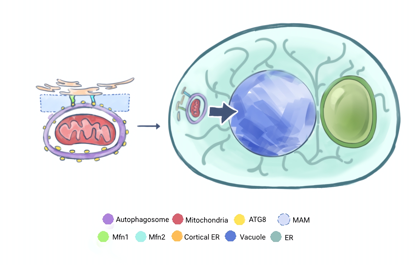
Here, Atg8 autophagosomes can form and traffic dysfunctional mitochondria material to the vacuole.
ER-Stress in ND and ER-phagy
The endoplasmic reticulum (ER) expands across much of the cell, creating membrane-contact sites with many fundamental organelles, such as the mitochondria, lysosome, and nucleus [25]. At these contact sites, lipid and ion transfer occurs between the two tethered organelles and a network system is established—important for protein synthesis [25,37]. The ER is also part of the secretory pathway, responsible for completing the translation of secretory proteins and trafficking them to the Golgi to be packaged. So, when there is ER stress, it means there are unfolded or misfolded protein aggregates in the ER, compromising ER function and thus the cell’s secretory pathway [28]. To date, there is more evidence of ER stress contributing to ND development, especially in AD and PD [26, 28]. So, we will now examine the ER stress response pathway ER-phagy, which is protective of neurons [8].
ER-phagy is an autophagic response to ER stress, and like lipophagy, it also utilizes NVJ. At NVJ, Atg39 assembles in the ER membrane and forms autophagosomes by binding to Atg8 [12], a core autophagy machinery protein. They do this by deriving autophagosomes from the nucleus, deforming the outer and inner nuclear membranes [10]. It is interesting to note that Atg39 is used in nucleophagy, further expanding the importance of NVJ [10, 12]. Since nutrient deficiency and proteostasis failure is common in ND, the overlap at NVJ for both nutrient and ER stress pathways may be targeted for investigation to see how it is altered under ND conditions. ER-phagy is shown to be protective of dopaminergic neurons against PD [8]. α-syn causes ER stress by accumulating in the ER, however proper ER-phagy activation will target α-syn rich areas, degrading α-syn and returning ER function [8]. How ER-phagy fails is the next question to ask, so we are curious if other autophagic pathways are intertwined with ER-phagy: For example, if the simultaneous activation of lipophagy and ER-phagy under PD conditions impacts α-syn clearance.
There is another ER-phagy receptor, Atg40, but this protein localizes along the cortical ER, which spreads far across the cell reaching the plasma membrane [9, 12]. Atg40 also recruits Atg8 to create autophagosomes, and will produce a curved structure of the ER regions selected for autophagy, allowing for autophagy of dysfunctional ER fragments [9]. This is similar to mitophagy which also relies on the cortical ER and Atg8 [13, 24].
Mitochondrial Stress in ND and Mitophagy
Mitochondria are key targets for ND researchers. Mitochondria are responsible for the cell’s energy production, and their dysfunction is involved in many NDs, such as Amyotrophic Lateral Sclerosis (ALS), Huntington’s Disease (HD), PD, and AD [18]. When mitochondria are damaged and non-functioning, it disrupts energy homeostasis and the cell must remove/recycle dysfunctional mitochondria through mitophagy, a selective autophagy [18]. There are several well-described mitophagy pathways, which may work in concert to compensate for defects in a parallel pathway [13, 37]. These pathways occur at MAMs, which are cortical ER-mitochondria contact sites, and recruits Atg8 for autophagosome formation [13, 24]. A measure of mitochondrial dysfunction in ND is the hyperactivation of mitophagy, as that would indicate more effort to degrade damaged mitochondria.
In AD, we see increased levels of mitophagy as mitochondrial dysfunction is exacerbated by Aβ toxicity [38]. Aβ overload caused a drop in the mitochondrial fusion proteins Mfn1 and Mfn2 [38], which suggests that mitophagy efficiency is reduced. Mfn1 and Mfn2 tethers the mitochondria to the MAM when mitophagy is initiated with AMP-activated protein kinase (AMPK) sensors on Mfn2 [24], so lower levels of these proteins would hinder mitophagy. Something similar happens in PD. In genetic PD, mutations for PINK1 and Parkin are the most frequent cause of early-onset PD, and these proteins work hand-in-hand to initiate mitophagy under mitochondrial depolarization [13, 39]. In non-genetic PD, overexpression of α-syn is shown to impair the mitochondrial electron transport chain’s complex I and reduce ATP levels [39]. In ND, there seems to be an increased demand for mitophagy, while there is a shortage of it.

nucleus-vacuole junction site. Then, autophagosomes carrying dysfunctional ER
material are trafficked to the vacuole for degradation.
While proteostasis and mitochondrial dysfunction are related, i.e., unfolded mitophagy receptors or lack of energy to synthesize proteins influence one another, they are independent pathways. We found no evidence of mitophagy occurring at the NVJ, and the ER-phagy receptors Atg39 and Atg40 do not influence mitophagy [12]. Both ER-phagy and mitophagy, however, require Atg8 [9, 24] for autophagosomes which emphasizes the importance of the core autophagy machinery proteins. Although these two pathways are independent of each other, they still rely on core autophagy machinery.
Conclusion and Outlook
Emerging evidence on lipophagy and ER-phagy mechanisms shows the importance of the NVJ to produce LDs and autophagosomes. The relationship between lipophagy and ER-phagy at NVJ is still unclear. It is unexplored if both pathways can simultaneously occur, or if the failure of one impacts the other. The NVJ shape, size, and protein crowding are key components for both lipophagy and ER-phagy. Raft formation modifies the vacuolar membrane [33], but there is little research done on the impact of rafts on other autophagic pathways, even though the pushing of the vacuolar membrane into “honey-combed” shapes [34] could impact the docking of other autophagosomes to the vacuole.
To date, there is little connection made between mitophagy and the cell responses to nutrient and ER stress, as mitophagy occurs at the cortical ER and is currently established as an independent pathway to ER-phagy [12]. The relationship between mitophagy and lipophagy is unclear, though there is substantial research on the impact of nutrient deprivation on mitochondria. For example, under nutrient deprivation, the mitochondria produces substantial amounts of reactive oxygen species (ROS) [40], which is toxic to the cell and another hallmark of ND and aging [41], suggesting mitochondrial stress. There must be other connections between different cell stress responses besides pathway.
ND has implications in nutrient, ER, and mitochondrial stress. In response, cells activate adaptive pathways to recover, but when cellular stress is too great for the cell’s capacity to respond, ND pathogenesis is set into motion. How to maintain autophagy efficiency or reduce cellular stress would ameliorate ND symptoms and are key topics of interest for ND researchers. In this review, we have highlighted the NVJ as an overlap between nutrient and ER stress response and shown the common reliance on the ER and core autophagy machinery in all three stress pathways. There are many more ND-relevant autophagic pathways to explore, such as nucleophagy and pexophagy, to further highlight the significance of autophagy for cellular homeostasis. Since multiple cell stressors are present in ND, it is key to examine the interconnective relationship between our autophagic pathways to understand how they may fail to protect us in ND pathogenesis.

About the Author: Brandon Fong
Brandon Fong is a junior at UC Davis majoring in Biochemistry and Molecular Biology, while minoring in Bioinformatics and Quantitative Biology. What sparked his interest in molecular biology is the fundamental question "How does a healthy cell become diseased?" His exploration led him to ask about dysfunctional autophagy, cell stress, and the Kaplan Lab. At the Kaplan Lab, Brandon investigates autophagic failure under a cell stress model. Understanding how autophagy fails could explain disease onset, as well as underlying cellular changes in aging. There are key high-interaction hubs of autophagy and reviewing their connection to various forms of autophagy can expand the perspective of cellular stress. Looking forward, Brandon aims to apply the lens of cell stress on the aging process, as well as translating aging research to geriatric care.
Author’s Note
There are many forms of autophagy, but it is surprising how much they intersect and overlap. Dysfunctional autophagy is ubiquitous in neurodegenerative disease, and a review on how different autophagy pathways relate to each other can provide inspiration for new experiments.
References
Vereinte Nationen, editor. 2023. Leaving no one behind in an ageing world. New York, NY: United Nations.
Hou Y, Dan X, Babbar M, Wei Y, Hasselbalch SG, Croteau DL, Bohr VA. 2019. Ageing as a risk factor for neurodegenerative disease. Nat Rev Neurol. 15(10):565–581. doi:10.1038/s41582-019-0244-7
Guo F, Liu X, Cai H, Le W. 2018. Autophagy in neurodegenerative diseases: pathogenesis and therapy. Brain Pathology. 28(1):3–13. doi:10.1111/bpa.12545
Da Silva PFL, Schumacher B. 2021. Principles of the Molecular and Cellular Mechanisms of Aging. Journal of Investigative Dermatology. 141(4):951–960. doi:10.1016/j.jid.2020.11.018
Twayana KS, Ravanan P. 2018. Eukaryotic cell survival mechanisms: Disease relevance and therapeutic intervention. Life Sciences. 205:73–90. doi:10.1016/j.lfs.2018.05.002
Guo J, Huang X, Dou L, Yan M, Shen T, Tang W, Li J. 2022. Aging and aging-related diseases: from molecular mechanisms to interventions and treatments. Sig Transduct Target Ther. 7(1):391. doi:10.1038/s41392-022-01251-0
Fleming A, Bourdenx M, Fujimaki M, Karabiyik C, Krause GJ, Lopez A, Martín-Segura A, Puri C, Scrivo A, Skidmore J, et al. 2022. The different autophagy degradation pathways and neurodegeneration. Neuron. 110(6):935–966. doi:10.1016/j.neuron.2022.01.017
Kim DY, Shin JY, Lee JE, Kim HN, Chung SJ, Yoo HS, Kim SJ, Cho HJ, Lee E-J, Nam SJ, et al. 2023. A selective ER-phagy exerts neuroprotective effects via modulation of α-synuclein clearance in parkinsonian models. Proc Natl Acad Sci U S A. 120(37):e2221929120. doi:10.1073/pnas.2221929120
Mochida K, Yamasaki A, Matoba K, Kirisako H, Noda NN, Nakatogawa H. 2020. Super-assembly of ER-phagy receptor Atg40 induces local ER remodeling at contacts with forming autophagosomal membranes. Nat Commun. 11(1):3306. doi:10.1038/s41467-020-17163-y
Mochida K, Otani T, Katsumata Y, Kirisako H, Kakuta C, Kotani T, Nakatogawa H. 2022. Atg39 links and deforms the outer and inner nuclear membranes in selective autophagy of the nucleus. J Cell Biol. 221(2):e202103178. doi:10.1083/jcb.202103178
Álvarez-Guerra I, Block E, Broeskamp F, Gabrijelčič S, Infant T, De Ory A, Habernig L, Andréasson C, Levine TP, Höög JL, Büttner S. 2024. LDO proteins and Vac8 form a vacuole-lipid droplet contact site to enable starvation-induced lipophagy in yeast. Developmental Cell. 59(6):759-775.e5. doi:10.1016/j.devcel.2024.01.014
Mochida K, Oikawa Y, Kimura Y, Kirisako H, Hirano H, Ohsumi Y, Nakatogawa H. 2015. Receptor-mediated selective autophagy degrades the endoplasmic reticulum and the nucleus. Nature. 522(7556):359–362. doi:10.1038/nature14506
Rühmkorf A, Harbauer AB. 2023. Role of Mitochondria-ER Contact Sites in Mitophagy. Biomolecules. 13(8):1198. doi:10.3390/biom13081198
López-Otín C, Blasco MA, Partridge L, Serrano M, Kroemer G. 2013. The Hallmarks of Aging. Cell. 153(6):1194–1217. doi:10.1016/j.cell.2013.05.039
Fluegge K. 2019. A model of lipid dysregulation and altered nutrient status in Alzheimer’s disease. A&D Transl Res & Clin Interv. 5(1):139–145. doi:10.1016/j.trci.2019.03.002
Liu GY, Sabatini DM. 2020. mTOR at the nexus of nutrition, growth, ageing and disease. Nat Rev Mol Cell Biol. 21(4):183–203. doi:10.1038/s41580-019-0199-y
Höhn A, Tramutola A, Cascella R. 2020. Proteostasis Failure in Neurodegenerative Diseases: Focus on Oxidative Stress. Oxidative Medicine and Cellular Longevity. 2020:1–21. doi:10.1155/2020/5497046
Bustamante-Barrientos FA, Luque-Campos N, Araya MJ, Lara-Barba E, De Solminihac J, Pradenas C, Molina L, Herrera-Luna Y, Utreras-Mendoza Y, Elizondo-Vega R, et al. 2023. Mitochondrial dysfunction in neurodegenerative disorders: Potential therapeutic application of mitochondrial transfer to central nervous system-residing cells. J Transl Med. 21(1):613. doi:10.1186/s12967-023-04493-w
Zhang S, Peng X, Yang S, Li X, Huang M, Wei S, Liu J, He G, Zheng H, Yang L, et al. 2022. The regulation, function, and role of lipophagy, a form of selective autophagy, in metabolic disorders. Cell Death Dis. 13(2):132. doi:10.1038/s41419-022-04593-3
Miller-Fleming L, Giorgini F, Outeiro TF. 2008. Yeast as a model for studying human neurodegenerative disorders. Biotechnol J. 3(3):325–338. doi:10.1002/biot.200700217
Xie Z, Klionsky DJ. 2007. Autophagosome formation: core machinery and adaptations. Nat Cell Biol. 9(10):1102–1109. doi:10.1038/ncb1007-1102
Kirkin V. 2020. History of the Selective Autophagy Research: How Did It Begin and Where Does It Stand Today? J Mol Biol. 432(1):3–27. doi:10.1016/j.jmb.2019.05.010
Dawaliby R, Mayer A. 2010. Microautophagy of the nucleus coincides with a vacuolar diffusion barrier at nuclear-vacuolar junctions. Mol Biol Cell. 21(23):4173–4183. doi:10.1091/mbc.E09-09-0782
Hu Y, Chen H, Zhang L, Lin X, Li X, Zhuang H, Fan H, Meng T, He Z, Huang H, et al. 2021. The AMPK-MFN2 axis regulates MAM dynamics and autophagy induced by energy stresses. Autophagy. 17(5):1142–1156. doi:10.1080/15548627.2020.1749490
Wu H, Carvalho P, Voeltz GK. 2018. Here, there, and everywhere: The importance of ER membrane contact sites. Science. 361(6401):eaan5835. doi:10.1126/science.aan5835
Singh R, Kaur N, Choubey V, Dhingra N, Kaur T. 2024. Endoplasmic reticulum stress and its role in various neurodegenerative diseases. Brain Research. 1826:148742. doi:10.1016/j.brainres.2023.148742
Colla E, Coune P, Liu Y, Pletnikova O, Troncoso JC, Iwatsubo T, Schneider BL, Lee MK. 2012. Endoplasmic Reticulum Stress Is Important for the Manifestations of α-Synucleinopathy In Vivo. J Neurosci. 32(10):3306–3320. doi:10.1523/JNEUROSCI.5367-11.2012
Ajoolabady A, Lindholm D, Ren J, Pratico D. 2022. ER stress and UPR in Alzheimer’s disease: mechanisms, pathogenesis, treatments. Cell Death Dis. 13(8):706. doi:10.1038/s41419-022-05153-5
Badaeva AV, Danilov AB, Clayton P, Moskalev AA, Karasev AV, Tarasevich AF, Vorobyeva YD, Novikov VN. 2023. Perspectives on Neuronutrition in Prevention and Treatment of Neurological Disorders. Nutrients. 15(11):2505. doi:10.3390/nu15112505
Lopes da Silva S, Vellas B, Elemans S, Luchsinger J, Kamphuis P, Yaffe K, Sijben J, Groenendijk M, Stijnen T. 2014. Plasma nutrient status of patients with Alzheimer’s disease: Systematic review and meta-analysis. Alzheimers Dement. 10(4):485–502. doi:10.1016/j.jalz.2013.05.1771
Boulos C, Yaghi N, El Hayeck R, Heraoui GN, Fakhoury-Sayegh N. 2019. Nutritional Risk Factors, Microbiota and Parkinson’s Disease: What Is the Current Evidence? Nutrients. 11(8):1896. doi:10.3390/nu11081896
Sheard JM. 2014. Malnutrition and Neurodegenerative Diseases. Curr Nutr Rep. 3(2):102–109. doi:10.1007/s13668-014-0078-2
Seo AY, Lau P-W, Feliciano D, Sengupta P, Gros MAL, Cinquin B, Larabell CA, Lippincott-Schwartz J. 2017. AMPK and vacuole-associated Atg14p orchestrate μ-lipophagy for energy production and long-term survival under glucose starvation. eLife. 6:e21690. doi:10.7554/eLife.21690
Toulmay A, Prinz WA. 2013. Direct imaging reveals stable, micrometer-scale lipid domains that segregate proteins in live cells. J Cell Biol. 202(1):35–44. doi:10.1083/jcb.20130103
Zhang A, Meng Y, Li Q, Liang Y. 2020. The endosomal sorting complex required for transport complex negatively regulates Erg6 degradation under specific glucose restriction conditions. Traffic. 21(7):488–502. doi:10.1111/tra.12732
Liao P-C, Garcia EJ, Tan G, Tsang CA, Pon LA. 2021. Roles for L o microdomains and ESCRT in ER stress-induced lipid droplet microautophagy in budding yeast. Mol Biol Cell. 32(22):br12. doi:10.1091/mbc.E21-04-0179
Perkins HT, Allan VJ, Waigh TA. 2021. Network organisation and the dynamics of tubules in the endoplasmic reticulum. Sci Rep. 11(1):16230. doi:10.1038/s41598-021-94901-2
De La Cueva M, Antequera D, Ordoñez-Gutierrez L, Wandosell F, Camins A, Carro E, Bartolome F. 2022. Amyloid-β impairs mitochondrial dynamics and autophagy in Alzheimer’s disease experimental models. Sci Rep. 12(1):10092. doi:10.1038/s41598-022-13683-3
Henrich MT, Oertel WH, Surmeier DJ, Geibl FF. 2023. Mitochondrial dysfunction in Parkinson’s disease – a key disease hallmark with therapeutic potential. Mol Neurodegeneration. 18(1):83. doi:10.1186/s13024-023-00676-7
Filomeni G, De Zio D, Cecconi F. 2015. Oxidative stress and autophagy: the clash between damage and metabolic needs. Cell Death Differ. 22(3):377–388. doi:10.1038/cdd.2014.150
Singh A, Kukreti R, Saso L, Kukreti S. 2019. Oxidative Stress: A Key Modulator in Neurodegenerative Diseases. Molecules. 24(8):1583. doi:10.3390/molecules24081583